Root Canal Treatment of Tooth #18:
Diagnosis: #18 Pulp Necrosis with Symptomatic Apical Periodontitis
Tooth was occluded to the upper denture. The patient reported pain and was not able to chew on the left side. Percussion and biting tests suggested that the pain was from the tooth #18. Tooth #18 is a necrotic tooth with secondary caries exposing the pulp. A periapical bone damage (i.e. periapical radiolucent lesion) was also noticed around the M and D roots of the tooth #18.

The pain resolved after the root canal treatment was completed on the tooth #18. At 1 year recall, the tooth remained asymptomatic with the healing of periapical bone.
Lower Molar with 5 canals: MB, Mid M, ML, DB, and DL canals
The patient presented with pain on a lower right tooth. Biting test reproduced the pain from the tooth #30. Root canal treatment was initiated previously by his dentist but the root canals were not visible and not located. Thus, the patient was referred to us for treatment. After the root canal treatment was completed on the tooth #30, the biting pain resolved completely.
Diagnosis: #30 Previously initiated therapy with symptomatic apical periodontitis

At 1 year recall, the tooth#30 remained asymptomatic with intact lamina dura and PDL space.
Nonsurgical retreatment of the tooth #3 due to an acute apical abscess from persisting infection from a missed/untreated MB2 root canal:
Diagnosis: #3 Previously treated tooth with acute apical abscess
The patient presented with a swelling on her upper right cheek and a buccal swelling next to the tooth #3. Root canal treatment was completed and a crown was placed on this tooth couple years ago. There was a periapical (PA) bone damage (i.e. periapical radiolucent lesion) around the MB root Of the tooth #3, where a periodontal pocket with 8 mm probing depth was noticed. Gingival recession was noticed with the exposure of the crown margin on the tooth #3 but there was no visible caries or leakage of the crown. Adjacent teeth, #2, 4, and 5 are asymptomatic vital teeth.
After a root canal access was made through an existing crown, a metal post and filling materials in the 3
previously treated root canals were removed. A fourth canal or MB2 was located. All 4 root canals were
cleaned completely and Ca(OH)2 was placed inside the root canals. At the second visit, the swelling and the periodontal pocket resolved completely. Thus the root canal treatment was completed and a composite filling was placed to close the access cavity.

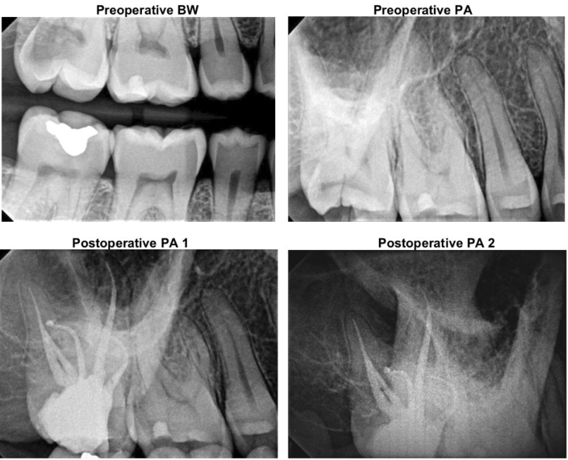
Upper Molar with 5 canals: MBI, MB2, OBI, DB2, and PA canals
Diagnosis: #2 Symptomatic Irreversible pulpitis with symptomatic apical periodontitis